4.2
Communication in the Nervous System
The blueprint we just described provides only a general idea of the nervous system's structure. Now let's turn to the details.
Types of Cells
Like the rest of your body, your brain is made of cells—two types of cells, in fact. Neurons, or nerve cells, are the brain's communication specialists, transmitting information to, from, and within the central nervous system. Glia, or glial cells (from the Greek for “glue”), hold the neurons in place.
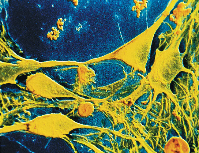
Neurons in the outer layers of the brain.
Although glia used to get much less attention than neurons, we now know that these cells are much more than just “glue.” They provide the neurons with nutrients, insulate them, help them grow, protect the brain from toxic agents, and remove cellular debris when neurons die. They also communicate chemically with each other and with neurons; without them, neurons could not function effectively. One kind of glial cell appears to give neurons the go-ahead to form connections and to start “talking” to each other (Ullian, Christopherson, & Barres, 2004). Another kind seems to act like the brain's electrician, identifying and trying to repair problems with the nerves' electrical systems (Graeber & Streit, 2010). And over time, glia help determine which neural connections get stronger or weaker, suggesting that they play a vital role in learning and memory (Fields, 2004).
It is the neurons, however, that are considered the building blocks of the nervous system, though in structure they are more like snowflakes than blocks, exquisitely delicate and differing from one another greatly in size and shape (see Figure4.3). In the giraffe, a neuron that runs from the spinal cord down the animal's hind leg may be 9 feet long! In the human brain, neurons are microscopic. Scientists believed for many years that the brain contains about 100 billion neurons and 10 times as many glia. But recent advances, which allow researchers to count individual cells, put the numbers much lower. An adult brain contains about 171 billion cells, about evenly divided between neurons and glia (Herculano-Houzel, 2009; Lent et al., 2012).
Figure 4.3
Different Kinds of Neurons
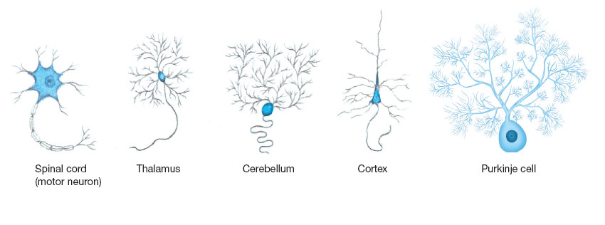
Neurons vary in size and shape, depending on their location and function. More than 200 types of neurons have been identified in mammals.
The Structure of the Neuron
As you can see in Figure4.4, a neuron has three main parts: dendrites, a cell body, and an axon. The dendrites look like the branches of a tree; indeed, the word dendrite means “little tree” in Greek. Dendrites act like antennas, receiving messages from as many as 10,000 other nerve cells and transmitting these messages toward the cell body. They also do some preliminary processing of those messages. The cell body is shaped roughly like a sphere or a pyramid; it includes the cell's nucleus, which contains genetic information (DNA) and controls the cell's growth and reproduction. The rest of the cell body contains the biochemical machinery for keeping the neuron alive and producing neurochemicals (which you will read about shortly). The axon (from the Greek for “axle”) is attached to the cell body and transmits messages away from the cell body to other neurons or to muscle or gland cells. Axons commonly divide at the end into branches called axon terminals. In adult human beings, axons vary from only four thousandths of an inch to a few feet in length. Dendrites and axons give each neuron a double role: As one researcher put it, a neuron is first a catcher, then a batter (Gazzaniga, 1988).
Figure4.4
The Structure of a Neuron
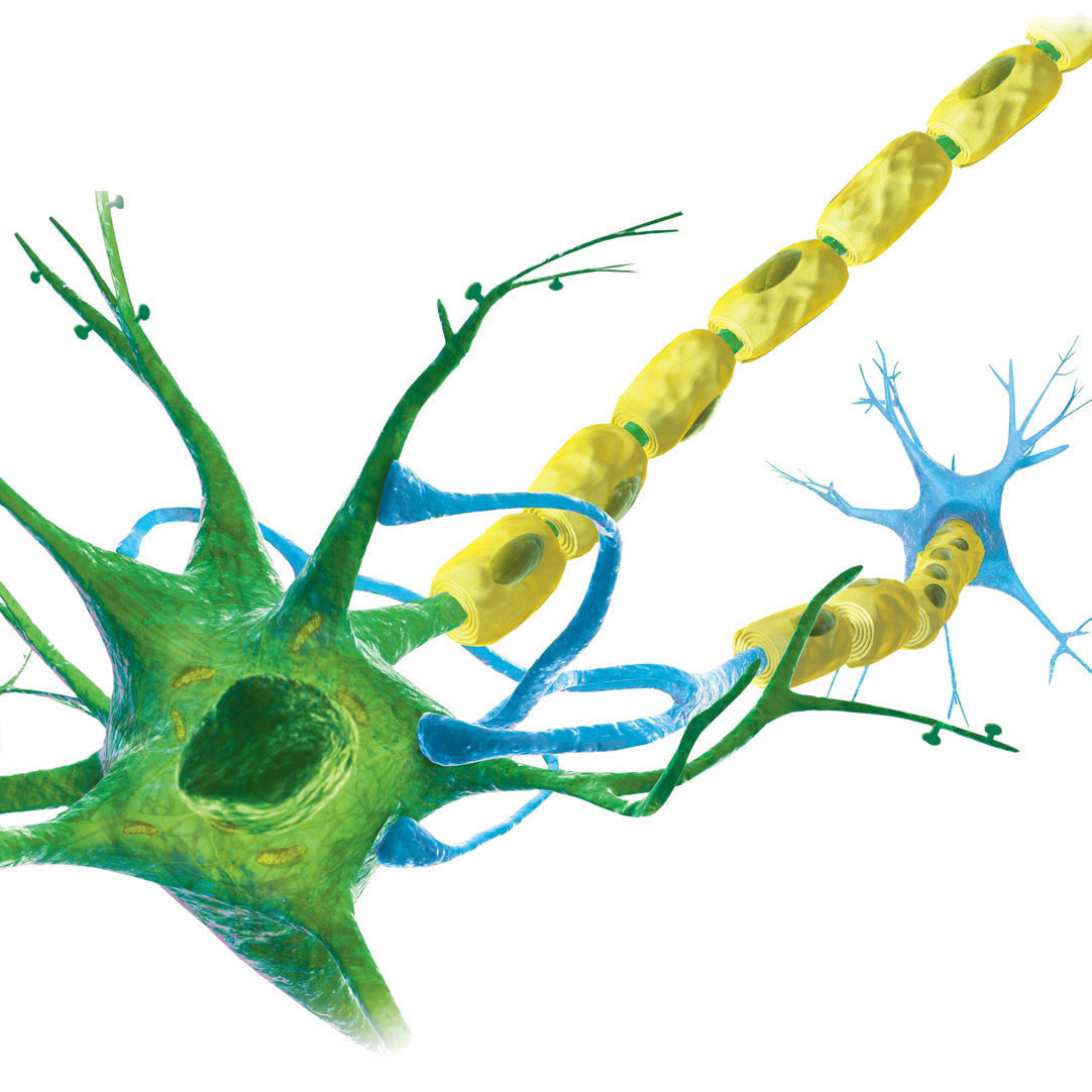
Incoming neural impulses are received by the dendrites of a neuron and are transmitted to the cell body. Outgoing signals pass along the axon to terminal branches.
Many axons, especially the larger ones, are insulated by a surrounding layer of fatty material called the myelin sheath, which in the central nervous system is made up of glial cells. Constrictions in this covering, called nodes, divide it into segments, which make it look a little like a string of link sausages (see Figure4.4 again). One purpose of the myelin sheath is to prevent signals in adjacent cells from interfering with each other. Another purpose, as we will see shortly, is to speed up the conduction of neural impulses. In individuals with multiple sclerosis, loss of myelin causes erratic nerve signals, leading to loss of sensation, weakness or paralysis, lack of coordination, or vision problems (Czeipel, Boddeke, & Copray, 2015). To learn more about neurons and how they work, watch the video The Basics: How the Brain Works 2.
Watch
The Basics: How the Brain Works 2
In the peripheral nervous system, the fibers of individual neurons (axons and sometimes dendrites) are collected together in bundles called nerves, rather like the lines in a telephone cable. The human body has 43 pairs of peripheral nerves; one nerve from each pair is on the left side of the body and the other is on the right. Most of these nerves enter or leave the spinal cord, but 12 pairs in the head, the cranial nerves, connect directly to the brain.
How Neurons Communicate
Neurons do not directly touch each other, end to end. Instead, they are separated by a minuscule space called the synaptic cleft, where the axon terminal of one neuron nearly touches a dendrite or the cell body of another. The entire site—the axon terminal, the cleft, and the covering membrane of the receiving dendrite or cell body—is called a synapse. Because a neuron's axon may have hundreds or even thousands of terminals, a single neuron may have synaptic connections with a great many others. As a result, the number of communication links in the nervous system runs into the trillions or perhaps even the quadrillions. To learn more about how these connections change and multiply over time, watch the video The Plastic Brain.
Watch
The Plastic Brain
Neurons speak to one another, or in some cases to muscles or glands, in an electrical and chemical language. The inside and outside of a neuron contain positively and negatively charged ions (electrically charged atoms). At rest, the neuron has a negative charge relative to the outside. But when it is stimulated, special “gates” in the cell's membrane open, allowing positively charged sodium ions to move from the outside to the inside, making the neuron less negative. If this change reaches a critical level, it briefly triggers an action potential, during which gates in the axon membrane allow even more positively charged sodium into the cell, causing it to become positively charged; as a result, the neuron “fires.” Then, positively charged potassium ions quickly move from within the axon to the outside, which returns the cell to its negatively charged resting state.
If an axon is unmyelinated, this process repeats stepwise down the axon like dominos falling over in a line. But in myelinated axons, the process is a little different. Conducting a neural impulse beneath the sheath is impossible, in part because sodium and potassium ions cannot cross the cell's membrane except at the breaks (nodes) between the myelin's “sausages.” Instead, the action potential “hops” from one node to the next. (More precisely, positively charged ions flow down the axon at a fast rate, causing regeneration of the action potential at each node.) This arrangement allows the impulse to travel faster than it could if the action potential had to be regenerated at every point along the axon. Nerve impulses travel more slowly in babies than in older children and adults because when babies are born, the myelin sheaths on their axons are not yet fully developed.
When a neural impulse reaches the axon terminal's button-like tip, it must get its message across the synaptic cleft to another cell. At this point, synaptic vesicles, tiny sacs in the tip of the axon terminal, open and release a few thousand molecules of a chemical substance called a neurotransmitter. Like sailors carrying a message from one island to another, these molecules then diffuse across the synaptic cleft (see Figure4.6).
Figure 4.6
Neurotransmitter Crossing a Synapse
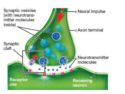
Neurotransmitter molecules are released into the synaptic cleft between two neurons from vesicles (chambers) in the transmitting neuron's axon terminal. The molecules then bind to receptor sites on the receiving neuron. As a result, the electrical state of the receiving neuron changes and the neuron becomes either more likely to fire an impulse or less so, depending on the type of neurotransmitter.
When they reach the other side, the neurotransmitter molecules bind briefly with receptor sites, special molecules in the membrane of the receiving neuron's dendrites (or sometimes cell body), fitting these sites much as a key fits a lock. Remember: The receiving neuron will be negatively charged because it is still at rest. Some neurotransmitters will cause a decrease in the negative charge. When the charge reaches a critical level, the neuron will fire. This is called an excitatory effect. Other neurotransmitters will cause an increase in the negative charge, making the neuron less likely to fire. This is called an inhibitory effect. Inhibition in the nervous system is essential. Without it, we could not sleep or coordinate our movements. Excitation of the nervous system would be overwhelming, producing convulsions.
What any given neuron does at any given moment depends on the net effect of all the messages being received from other neurons. Only when the cell's voltage reaches a certain threshold will it fire. Thousands of messages, both excitatory and inhibitory, may be coming into the cell, and the receiving neuron must essentially average them. The message that reaches a final destination depends on the rate at which individual neurons are firing, how many are firing, what types of neurons are firing, where the neurons are located, and the degree of synchrony among different neurons. It does not depend on how strongly the individual neurons are firing, however, because a neuron always either fires or doesn't. The firing of a neuron is an all-or-none event, like turning on a light switch. For a better idea of how neurons communicate with one another, watch the video The Basics: How the Brain Works 3.
Watch
The Basics: How the Brain Works 3
Chemical Messengers in the Nervous System
The nervous system “house” would remain forever dark and lifeless without chemical couriers and helpers to carry messages from room to room. We will consider three classes of chemicals: neurotransmitters, hormones, and neuromodulators.
Neurotransmitters: Versatile Couriers As we have seen, neurotransmitters make it possible for one neuron to excite or inhibit another. Neurotransmitters exist not only in the brain but also in the spinal cord, the peripheral nerves, and certain glands. Through their effects on specific nerve circuits, these substances control everything your brain does. The nature of the effect depends on the level of the neurotransmitter, its location, and the type of receptor it binds with. Here we will discuss just a few of the better-understood neurotransmitters and some of their known or suspected effects.
Four neurotransmitters each travel a particular path through parts of the brain, like following a bus route:
Serotonin affects neurons involved in sleep, appetite, sensory perception, temperature regulation, pain suppression, and mood.
Dopamine affects neurons involved in voluntary movement, attention, learning, memory, emotion, pleasure and reward, and possibly responses to novelty.
Acetylcholine affects neurons involved in muscle action, arousal, vigilance, memory, and emotion.
Norepinephrine affects neurons involved in increased heart rate and the slowing of intestinal activity during stress, and neurons involved in learning, memory, dreaming, waking from sleep, and emotion.
Two other common neurotransmitters are distributed throughout the entire brain:
GABA(gamma aminobutyric acid) is the major inhibitory neurotransmitter in the brain.
Glutamate is the major excitatory neurotransmitter in the brain; it is released by about 90 percent of the brain's neurons.
Harmful effects can occur when neurotransmitter levels are too high or too low. Abnormal GABA levels have been implicated in sleep and eating disorders and in convulsive disorders, including epilepsy. People with Alzheimer's disease lose brain cells responsible for producing acetylcholine and other neurotransmitters, and these deficits help account for their devastating memory problems. A loss of cells that produce dopamine is responsible for the tremors and rigidity of Parkinson's disease. In multiple sclerosis, immune cells overproduce glutamate, which damages or kills glial cells that normally make myelin. The video Neurotransmitters will show you more about how one of these important chemical messengers, dopamine, works.
Watch
Neurotransmitters
We want to warn you, however, that pinning down the relationship between neurotransmitter abnormalities and behavioral or physical abnormalities is extremely tricky. Each neurotransmitter plays multiple roles, and the functions of different substances often overlap. Furthermore, it is always possible that something about a disorder leads to abnormal neurotransmitter levels instead of the other way around. Correlation is not the same thing as causation. Finally, although drugs that boost or decrease levels of particular neurotransmitters are sometimes effective in treating certain mental disorders, this fact does not necessarily mean that abnormal neurotransmitter levels cause the disorders. After all, aspirin can relieve a headache, but headaches are not caused by a lack of aspirin!
Many of us regularly ingest things that affect our own neurotransmitters. Most recreational drugs produce their effects by blocking or enhancing the actions of neurotransmitters, and so do some herbal remedies. St. John's wort, which is often taken for depression, prevents the cells that release serotonin from reabsorbing excess molecules that have remained in the synaptic cleft; as a result, serotonin levels rise. Many people do not realize that such remedies, because they affect the nervous system's biochemistry, can interact with other medications and can be harmful in high doses. Even ordinary foods can influence the availability of neurotransmitters in the brain. Serotonin levels will decrease after a protein-rich meal (dairy products, meat, fish, and poultry) and increase after a high-carbohydrate, low-protein meal, which is why you may feel calm or lethargic after downing a big bowl of pasta (Spring, Chiodo, & Bowen, 1987). But the path between the bowl and the brain is complicated: If you're looking for brain food, you are most likely to find it in a well-balanced diet. Watch the video Your Brain on Drugs to learn more about how various substances can affect the brain.
Watch
Your Brain on Drugs
Hormones: Long-Distance MessengersHormones, which make up the second class of chemical messengers, are produced primarily in endocrine glands, such as the pancreas, ovaries, testes, and adrenal glands. Hormones are released directly into the bloodstream, which carries them to organs and cells that may be far from their point of origin. Hormones have dozens of jobs, from promoting bodily growth to aiding digestion to regulating metabolism. Receptors for hormones exist throughout the body, including the brain. Because hormones are released into the bloodstream, their effects are widespread.
Neurotransmitters and hormones are not always chemically distinct because nature has been efficient, giving some substances more than one role. For instance, norepinephrine may be considered either a neurotransmitter or a hormone, depending on where it is located and what function it is performing.

The following hormones, among others, are of particular interest to research psychologists and neuroscientists:
Melatonin, which is secreted by the pineal gland deep within the brain, helps to regulate daily biological rhythms and promotes sleep.
Oxytocin, which is secreted by another small gland in the brain, the pituitary gland, enhances uterine contractions during childbirth and facilitates the ejection of milk during nursing. Along with another hormone, vasopressin, oxytocin contributes to relationships in both sexes by promoting attachment and trust.
Adrenal hormones, which are produced by the adrenal glands (organs that are perched right above the kidneys), are involved in emotion and stress. These hormones also rise in response to other conditions, such as heat, cold, pain, injury, burns, and physical exercise, and in response to some drugs, such as caffeine and nicotine. The outer part of each adrenal gland produces cortisol, which increases blood sugar levels and boosts energy. The inner part produces epinephrine (commonly known as adrenaline) and norepinephrine. When adrenal hormones are released in your body, activated by the sympathetic nervous system, they increase your arousal level and prepare you for action. Adrenal hormones also enhance memory.
Sex hormones, which are secreted by tissue in the gonads (testes in men, ovaries in women) and also by the adrenal glands, include three main types, all occurring in both sexes but in differing amounts and proportions in males and females after puberty. Androgens (the most important of which is testosterone) are masculinizing hormones produced mainly in the testes but also in the ovaries and the adrenal glands. Androgens set in motion the physical changes males experience at puberty, including a deepened voice and facial and chest hair, and they cause pubic and underarm hair to develop in both sexes. Testosterone also influences sexual arousal in both sexes. Estrogens are feminizing hormones that bring on physical changes in females at puberty, such as breast development and the onset of menstruation, and that influence the course of the menstrual cycle. Progesterone contributes to the growth and maintenance of the uterine lining in preparation for a fertilized egg, among other functions. Estrogens and progesterone are produced mainly in the ovaries but also in the testes and the adrenal glands.
Sex hormones act on the brain to guide sexual behavior, but they are also involved in behavior not linked to sex or reproduction. The body's natural estrogen in both sexes is thought to enhance learning and memory by promoting the formation of synaptic connections in certain areas of the brain and by indirectly increasing the production of acetylcholine (Gibbs, 2010; Lee & McEwen, 2001; Sherwin, 1998). However, the common belief that fluctuating levels of estrogen and progesterone make most women “emotional” before menstruation has not been borne out by research.
Neuromodulators: The Brain's Volume Control The brain is awash in thousands of other chemicals that affect how neurons and neurotransmitters function (Brezina, 2010). Because these chemicals modulate (vary the strength of) neural functions, they are called neuromodulators. One neuromodulator, the serotonin transporter, is a protein that acts like a garbage collector, picking up serotonin from the synaptic cleft after it has been released and transporting it back to the sending neuron for recycling. In that way, it controls the amount of serotonin that is available in the brain.
Endorphins are an intriguing group of chemicals known technically as endogenous opioid peptides. Endorphins have effects similar to those of natural opiates such as heroin; that is, they reduce pain and promote pleasure. They are also thought to play a role in appetite, sexual activity, blood pressure, mood, learning, and memory. Some endorphins function as neurotransmitters, but most of them act primarily as neuromodulators, by limiting or prolonging the effects of neurotransmitters.
Endorphin levels shoot up when an animal or a person is afraid or under stress. This is no accident; by making pain bearable in such situations, endorphins give a species an evolutionary advantage. When an organism is threatened, it needs to do something fast. But pain can interfere with action: A mouse that pauses to lick a wounded paw may become a cat's dinner; a soldier who is overcome by an injury may never get off the battlefield. Of course, the body's built-in system of counteracting pain is only partly successful, especially when painful stimulation is prolonged.
A link also exists between endorphins and human attachment. Research with animals suggests that in infancy, contact with the mother stimulates the flow of endorphins, which strengthens the infant's bond with her. Some researchers now think that this “endorphin rush” also occurs in the early stages of passionate love between adults, accounting for the feeling of euphoria that “falling” for someone creates (Diamond, 2004).