4.4
A Tour Through the Brain
Most modern brain theories assume that different brain parts perform different (though greatly overlapping) tasks. This concept, known as localization of function, goes back at least to Joseph Gall (1758–1828), an Austrian anatomist who thought that personality traits were reflected in the development of specific areas of the brain. Gall's theory of phrenology was completely wrongheaded (so to speak), but his general notion of specialization in the brain had merit.
To learn about what the major brain structures do, let's take an imaginary stroll through the brain. Pretend that you have shrunk to a microscopic size and that you are wending your way through the “soul's frail dwelling house,” starting at the lower part, just above the spine. Figure4.10 shows the major structures we will encounter along our tour; you may want to refer to it as we proceed. But keep in mind that any activity—feeling an emotion, having a thought, performing a task—involves many different structures working together. Our description, therefore, is a simplification.
Figure4.10
Major Structures of the Human Brain
This cross section depicts the brain as if it were split down the middle and shows the structures described in the text
The Brain Stem and Cerebellum
We begin at the base of the skull with the brain stem, which began to evolve some 500 million years ago in segmented worms. The brain stem looks like a stalk rising out of the spinal cord. Pathways to and from upper areas of the brain pass through its two main structures: the medulla and the pons. The pons is involved in (among other things) sleeping, waking, and dreaming. The medulla is responsible for bodily functions that do not have to be consciously willed, such as breathing and heart rate. Hanging has long been used as a method of execution because when it breaks the neck, nerve pathways from the medulla are severed, stopping respiration.
Extending upward from the core of the brain stem is the reticular activating system (RAS). This dense network of neurons, which extends above the brain stem into the center of the brain and has connections with areas that are higher up, screens incoming information and arouses the higher centers when something happens that demands their attention. Without the RAS, we could not be alert or perhaps even conscious.
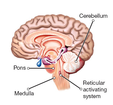
Standing atop the brain stem and looking toward the back part of the brain, we see a structure about the size of a small fist. It is the cerebellum, or “lesser brain,” which contributes to a sense of balance and coordinates the muscles so that movement is smooth and precise. If your cerebellum were damaged, you would probably become exceedingly clumsy and uncoordinated. You might have trouble using a pencil, threading a needle, or even walking. In addition, this structure is involved in remembering simple skills and acquired reflexes (Daum & Schugens, 1996; Krupa, Thompson, & Thompson, 1993). But the cerebellum, which was once considered just a motor center, is not as “lesser” as its name implies; it is involved in cognitive and emotional learning (Durisko & Fiez, 2010; Mariën et al., 2014).
The Thalamus
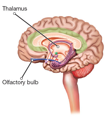
Deep in the brain's interior, roughly at its center, we can see the thalamus, the sensory relay station of the brain. As sensory messages come into the brain—about the sight of a sunset, the sound of a siren, the feel of a fly landing on your arm—the thalamus directs them to higher areas in charge of vision, sound, or touch. The only sense that completely bypasses the thalamus is the sense of smell, which has its own private switching station, the olfactory bulb. The olfactory bulb lies near areas involved in emotion, which may be why particular odors, such as the smell of fresh laundry or a steaming bowl of chicken soup, often rekindle vivid memories.
The Hypothalamus and the Pituitary Gland
Beneath the thalamus sits a structure called the hypothalamus (hypo means “under”). It is the body's boss, constantly monitoring the body's current state and issuing instructions to help the body maintain a steady state called homeostasis. It is involved in basic survival drives associated with the “four Fs”—feeding, fighting, fleeing, and . . . sex. It regulates body temperature by triggering sweating or shivering, and it controls the complex operations of the autonomic nervous system. It also contains the biological clock that controls the body's daily rhythms.
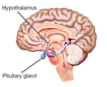
Hanging down from the hypothalamus, connected to it by a short stalk, is a cherry-sized endocrine gland called the pituitary gland, mentioned earlier in our discussion of hormones. The pituitary is often called the body's “master gland” because the hormones it secretes affect many other endocrine glands. The master, however, is really only a supervisor. The true boss is the hypothalamus, which sends chemicals to the pituitary that tell it when to “talk” to the other endocrine glands. The pituitary, in turn, sends hormonal messages out to these glands.
The hypothalamus and several other loosely interconnected structures have often been considered part of the limbic system; the term is from the Latin for “border,” and these structures were thought to form a border between the “higher” and “lower” parts of the brain. Structures in this region are heavily involved in emotions that we share with other animals, such as rage and fear, so the region is also sometimes called “the emotional brain.” But researchers now know that these structures also have other functions, and that parts of the brain outside of the old limbic system are involved in emotion. As a result, the term limbic system has been going out of favor.
The Amygdala
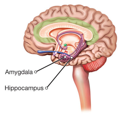
The amygdala (from the ancient Greek word for “almond”) is responsible for evaluating sensory information, quickly determining its emotional importance, and contributing to the initial decision to approach or withdraw from a person or situation. Some people describe the amygdala as the brain's “fear center,” but it is more than that. Your amygdala assesses stimuli for their fit with your current psychological state and even your core personality traits, responding to positive, negative, or even just plain interesting stimuli accordingly. It does so by working with higher brain areas to regulate your response. If you're extroverted, your amygdala responds more actively to photographs of happy people than it does if you are shy, and when you're hungry, your amygdala responds to food (Cunningham & Brosch, 2012). The amygdala also plays a role in mediating anxiety and depression and in forming and retrieving emotional memories.
The Hippocampus
The shape of the hippocampus must have reminded someone of a sea horse, for in Latin that is what its name means. The structure compares sensory information with what the brain has learned about the world, and when a match occurs, the hippocampus tells the reticular activating system to “cool it.” There's no need for neural alarm bells to go off every time a car goes by, a bird chirps, or you feel your saliva trickling down the back of your throat.
The hippocampus is also the brain's primary memory structure, and indeed has often been called the “gateway to memory.” It enables us to take in and combine different components of experiences—sights, sounds, and feelings—and bind them together into one “memory,” although the individual components may ultimately be stored in various parts of the cerebral cortex, which we will discuss shortly. When you recall meeting someone yesterday, various aspects of the memory—information about the person's greeting, tone of voice, appearance, and location—are probably stored in different locations in the cortex. But without the hippocampus, the information would never get to these destinations. This structure is also involved in the retrieval of information during recall.
We know about the central role the hippocampus plays in memory in part from research on patients whose brain damage produced severe memory problems. The most famous of these was Henry Molaison, known to the scientific community as H. M. When Henry was a child, he hit his head in a collision with a cyclist, and soon thereafter, possibly as a result, he began having seizures. By the time he was an adult, his blackouts and convulsions were preventing him from holding a job. He was referred to a surgeon who made the bold suggestion to remove Henry's hippocampus and some portion of his temporal lobes. Although the operation controlled Henry's epilepsy, it created a new problem for him: Henry could no longer form new memories for facts and events. He could remember his childhood, learn new skills, and carry on an intelligent conversation. But if you met him yesterday, he would not know you today. A lot of what psychological scientists know about the hippocampus and about memory is thanks to H. M.
The Cerebrum
At this point in our tour, the largest part of the brain still looms above us. It is the cauliflower-like cerebrum, where the higher forms of thinking take place. The complexity of the human brain's circuitry far exceeds that of any computer in existence, and much of its most complicated wiring is packed into this structure. Compared to many other creatures, we humans may be ungainly, feeble, and thin-skinned, but our well-developed cerebrum enables us to overcome these limitations and creatively control our environment (and, some would say, to mess it up).
The cerebrum is divided into two separate halves, or cerebral hemispheres, connected by a large band of fibers called the corpus callosum. In general, the right hemisphere is in charge of the left side of the body, and the left hemisphere is in charge of the right side of the body. As we will see shortly, the two hemispheres also have somewhat different tasks and talents, a phenomenon known as lateralization.
The Cerebral Cortex
Working our way right up through the top of the brain, we find that the cerebrum is covered by several thin layers of densely packed cells known collectively as the cerebral cortex. Cell bodies in the cortex, as in many other parts of the brain, produce a grayish tissue, hence the term gray matter. In other parts of the brain (and in the rest of the nervous system), long, myelin-covered axons prevail, providing the brain's white matter. Although the cortex is only about 3 millimeters (1/8 inch) thick, it contains almost three-fourths of all the cells in the human brain. The cortex has many deep crevasses and wrinkles, which enable it to contain its billions of neurons without requiring us to have the heads of giants—heads that would be too big to permit us to be born. In other mammals, which have fewer neurons, the cortex is less crumpled; in rats, it is quite smooth.
Lobes of the Cortex In each cerebral hemisphere, deep fissures divide the cortex into four distinct regions, or lobes (see Figure4.11):
Figure4.11
Lobes of the Cerebrum
Deep fissures divide the cortex of each cerebral hemisphere into four regions.
The occipital lobes (from the Latin for “in back of the head”) are at the lower back part of the brain. Among other things, they contain the visual cortex, where visual signals are processed. Damage to the visual cortex can cause impaired visual recognition or blindness.
The parietal lobes (from the Latin for “pertaining to walls”) are at the top of the brain. They contain the somatosensory cortex, which receives information about pressure, pain, touch, and temperature from all over the body. The areas of the somatosensory cortex that receive signals from the hands and the face are disproportionately large because these body parts are particularly sensitive. Parts of the parietal lobes are also involved in attention and awareness of spatial relationships.
The temporal lobes (from the Latin for “pertaining to the temples”) are at the sides of the brain, just above the ears and behind the temples. They are involved in memory, perception, and emotion, and they contain the auditory cortex, which processes sounds. An area of the left temporal lobe known as Wernicke's area is involved in language comprehension.
The frontal lobes, as their name indicates, are located toward the front of the brain, just under the skull in the area of the forehead. They contain the motor cortex, which issues orders to the 600 muscles of the body that produce voluntary movement. In the left frontal lobe, a region known as Broca's area handles speech production. During short-term memory tasks, areas in the frontal lobes are especially active. The frontal lobes are also involved in emotion and in the ability to make plans, think creatively, and take initiative.
Because the lobes of the cerebral cortex have different functions, they tend to respond differently when directly stimulated with tiny electrodes during brain surgery. (The brain does not feel anything when directly stimulated, so a patient can be awake during the operation.) If a surgeon touches the somatosensory cortex in the parietal lobes, a patient might feel a tingling in the skin or a sense of being gently touched. If the visual cortex in the occipital lobes were electrically stimulated, he or she might report a flash of light or swirls of color. And, eerily, many areas of the cortex, when stimulated, would produce no obvious response or sensation. These “silent” areas are sometimes called the association cortex because they are involved in higher mental processes. To review the major structures of the brain, watch the video How the Brain Works Part 4.
Watch
How the Brain Works 4
The Prefrontal Cortex The most forward part of the frontal lobes is the prefrontal cortex. This area barely exists in mice and rats and takes up only 3.5 percent of the cerebral cortex in cats and about 7 percent in dogs, but it accounts for approximately one-third of the entire cortex in human beings. It is the most recently evolved part of our brains, and is associated with such complex abilities as reasoning, decision making, and planning.
Scientists have long known that the frontal lobes, and the prefrontal cortex in particular, must also have something to do with personality. The first clue appeared in 1848, when a bizarre accident drove an inch-thick, 3½-foot-long iron rod clear through the head of a young railroad worker named Phineas Gage. The rod (which is still on display at the Harvard University Medical School library, along with Gage's skull) entered beneath the left eye and exited through the top of the head, destroying much of the prefrontal cortex (H. Damasio et al., 1994; Van Horn et al., 2012). Miraculously, Gage survived this trauma and, by most accounts, he retained the ability to speak, think, and remember. But his friends complained that he was “no longer Gage.” In a sort of Jekyll-and-Hyde transformation, he had changed from a mild-mannered, friendly, efficient worker into a foul-mouthed, ill-tempered, undependable lout who could not hold a steady job or stick to a plan. His employers had to let him go, and he was reduced to exhibiting himself as a circus attraction.
There is some controversy about the details of this sad incident, but many other cases of brain injury, whether from stroke or trauma, support the conclusion that most scientists draw from the Gage case: Parts of the frontal lobes are involved in social judgment, rational decision making, and the ability to set goals and to make and carry through plans. Like Gage, people with damage in these areas sometimes mismanage their finances, lose their jobs, and abandon their friends. Interestingly, the mental deficits that characterize damage to these areas are accompanied by a flattening-out of emotion and feeling, which suggests that normal emotions are necessary for everyday reasoning and the ability to learn from mistakes (A. Damasio, 2003; H. Damasio et al., 1994).
The frontal lobes also govern the ability to do a series of tasks in the proper sequence and to stop doing them at the proper time. Pioneering Soviet psychologist Alexander Luria (1980) studied many cases in which damage to the frontal lobes disrupted these abilities. One man kept trying to light a match after it was already lit. Another planed a piece of wood in the hospital carpentry shop until it was gone and then went on to plane the workbench! Watch The Prefrontal Cortex to learn more about how this important area of the brain influences behavior.
Watch
Thinking Like a Psychologist: The Prefrontal Cortex
Review4.1 summarizes the major parts of the brain that we have discussed and their primary functions.
Review4.1
Major Brain Structures and Functions