4.5
The Two Hemispheres of the Brain
We have seen that the cerebrum is divided into two hemispheres that control opposite sides of the body. Although similar in structure, these hemispheres have somewhat separate talents, or areas of specialization. Hemispheric specialization is especially apparent in patients who have suffered brain damage, usually as a result of a stroke. Those with left-hemisphere damage may lose the ability to speak or understand language, whereas those with right-hemisphere damage rarely do. A French neurologist named Paul Broca (whose name lives on in the term Broca's area) first made this observation in 1861, and many behavioral and cognitive difficulties associated with left- or right-hemisphere damage have been documented since then. Patients with left-hemisphere damage may have difficulties with reading, identifying objects, making symbolic gestures or pantomimes, and describing events in the correct order. For their part, patients with right-hemisphere damage may have difficulty identifying faces, interpreting emotional expressions in a face or voice, or understanding music or art. They may get lost easily, even in their own homes.
Split Brains: A House Divided
Some of the most interesting findings about hemispheric specialization come from people known as “split-brain patients.” In a normal brain, the two hemispheres of the cortex communicate with one another across the corpus callosum, the bundle of fibers that connects them. Whatever happens in one side of the brain is instantly transferred to the other side. What would happen, though, if the two sides were cut off from one another?
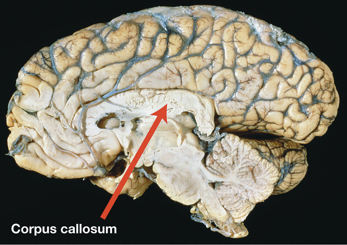
A cross section of a human brain, showing the corpus callosum.
In 1953, Ronald E. Myers and Roger W. Sperry took the first step toward answering this question by severing the corpus callosum in cats. They also cut parts of the nerves leading from the eyes to the brain. Normally, each eye transmits messages to both sides of the brain. After this procedure, a cat's left eye sent information only to the left hemisphere and its right eye sent information only to the right hemisphere.
At first, the cats did not seem to be affected much by this drastic operation. But Myers and Sperry showed that something profound had happened. They trained the cats to perform tasks with one eye blindfolded; a cat might learn to push a panel with a square on it to get food but ignore a panel with a circle. Then the researchers switched the blindfold to the cat's other eye and tested the animal again. Now the cats behaved as if they had never learned the trick. Apparently, one side of the brain did not know what the other side was doing; it was as if the animals had two brains in one body. Later studies confirmed this result with other species, including monkeys (Sperry, 1964).
In the animal studies, ordinary behavior, such as eating and walking, remained normal. In the early 1960s, a team of surgeons decided to try cutting the corpus callosum in patients with debilitating, uncontrollable epilepsy. In severe forms of this disease, disorganized electrical activity spreads from an injured area to other parts of the brain. The surgeons reasoned that cutting the connection between the two halves of the brain might stop the spread of electrical activity from one side to the other. The surgery was done, of course, for the sake of the patients, who were desperate. But there was a bonus for scientists, who would be able to find out what each cerebral hemisphere can do when it is quite literally cut off from the other.
The results of this split-brain surgery generally proved successful. Seizures were reduced and sometimes disappeared completely. In their daily lives, split-brain patients did not seem much affected by the fact that the two hemispheres were incommunicado. Their personalities and intelligence remained intact; they could walk, talk, and lead fairly normal lives. Apparently, connections in the undivided deeper parts of the brain kept body movements and other functions normal. The two functioning hemispheres were each doing their own job; they just couldn't communicate with each other. But in a series of ingenious studies, Sperry and his colleagues (and later other researchers) showed that perception and memory had been affected, just as they had been in the earlier animal research. Sperry won a Nobel Prize for his work.
To understand this research, you must know how nerves connect the eyes to the brain. (The human patients, unlike Myers and Sperry's cats, did not have these nerves cut.) If you look straight ahead, everything in the left side of the scene before you—the visual field—goes to the right half of your brain, and everything in the right side of the scene goes to the left half of your brain. This is true for both eyes (see Figure4.12).
Figure 4.12
Visual Pathways
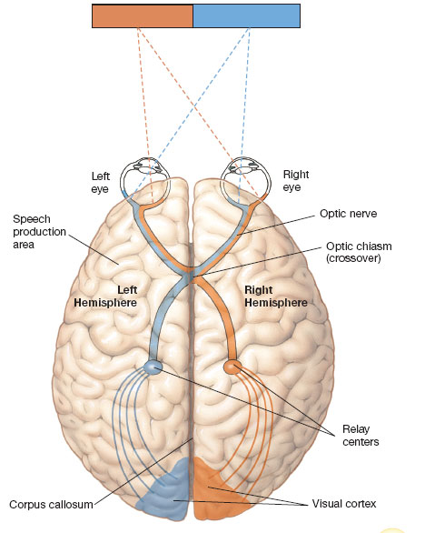
Each cerebral hemisphere receives information from the eyes about the opposite side of the visual field. Thus, if you stare directly at the corner of a room, everything to the left of the juncture is represented in your right hemisphere, and vice versa. This is so because half the axons in each optic nerve cross over (at the optic chiasm) to the opposite side of the brain. Normally, each hemisphere immediately shares its information with the other one, but in split-brain patients, severing the corpus callosum prevents such communication.
The procedure was to present information only to one or the other side of the patients' brains. In one early study, the researchers took photographs of different faces, cut them in two, and pasted different halves together (Levy, Trevarthen, & Sperry, 1972). The reconstructed photographs were then presented on slides. The person was told to stare at a dot in the middle of the screen, so that half of the image fell to the left of this point and half to the right. Each image was flashed so quickly that the person had no time to move his or her eyes. When the patients were asked to say what they had seen, they named the person in the right part of the image (which would be the little boy in Figure4.13). But when they were asked to point with their left hands to the face they had seen, they chose the person in the left side of the image (the mustached man in the figure). Furthermore, they claimed they had noticed nothing unusual about the original photographs! Each side of the brain saw a different half image and automatically filled in the missing part. Neither side knew what the other side had seen.
Figure4.13
A Split-Brain Experiment
Why did the patients name one side of the picture but point to the other? When the patient responded with speech, it was the left side of the brain, which usually controls speech, doing the talking. And because the left side of the brain had only seen the right side of the image, that was the face it saw. When the person pointed with the left hand, which is controlled by the right side of the brain, the right hemisphere was giving its version of what it had seen.
In another study, the researchers presented slides of ordinary objects and then suddenly flashed a slide of a nude woman. Both sides of the brain were amused, but because only the left side had speech, the two sides responded differently. When the picture was flashed to one woman's left hemisphere, she laughed and identified it as a nude. When it was flashed to her right hemisphere, she said nothing but began to chuckle. Asked what she was laughing at, she said, “I don't know . . . nothing . . . oh—that funny machine.” The right hemisphere could not describe what it had seen, but it reacted emotionally just the same, and the talking left hemisphere was compelled to come up with a reasonable explanation for the laughter (Gazzaniga, 1967). Indeed, Michael Gazzaniga (1989), a leading psychological scientist who conducted this and other split-brain research, has called the left hemisphere an “interpreter” because one of its major roles is to continually provide a reasonable (though not always accurate) story to explain our thoughts, feelings, and behaviors. As another neuroscientist put it, the left hemisphere is the brain's “spin doctor” (Broks, 2004).

Have a righthanded friend tap on a sheet of paper with a pencil held in the right hand for 1 minute. Then have the person do the same with the left hand, using a fresh sheet of paper. Finally, repeat the procedure, having the person talk at the same time as tapping. For most people, talking will decrease the rate of tapping—but more for the right hand than for the left, probably because both activities involve the same hemisphere (the left one), and there is competition between them. (Lefthanded people vary more in terms of which hemisphere is dominant for language, so the results for them will be more variable.)
The Two Hemispheres: Allies or Opposites?
The split-brain operation is still being performed, although more rarely, now that better medications are available to treat epilepsy. However, there is a limit to the conclusions we can draw from split-brain patients. The fact that their seizures were severe enough to warrant such dramatic surgery indicates that they probably suffered neurological damage at some point, which could have affected the results.
Fortunately, research on left–right differences has also been done with people whose brains are intact (Hugdahl & Westerhausen, 2010; Prete et al., 2015). If a researcher flashes an image very quickly to the right side of your visual field, it will arrive first in your left visual cortex. Thanks to your corpus callosum, the image will then quickly transfer over to your right visual cortex. But your left visual areas will have had a small head start, and the difference in timing gives the investigator critical information. People read words faster if the words are flashed to the right visual field because those words go directly to the left hemisphere, which is specialized for reading. In contrast, people are better at identifying facial expressions that are flashed to the left visual field because those faces go directly to the right hemisphere, which typically shows an advantage for processing facial emotion (Abbott et al., 2014). Similarly, researchers can present different sounds to your two ears, the one in the left ear going to your right auditory cortex and the one in the right ear going to the left auditory cortex. These studies show that although the left hemisphere is specialized for processing words, the right hemisphere is specialized for processing the tone of voice in which the words are spoken (Grimshaw et al., 2003).
Although early researchers often spoke of the left hemisphere as dominant, especially because of its linguistic and analytic talents, over the years it has become clear that the right hemisphere is far from stupid or passive. It is superior not only at recognizing facial expressions but also at handling problems requiring spatial–visual ability, the ability you use to read a map or follow a dress pattern. It is active during the creation and appreciation of art and music. It recognizes nonverbal sounds, such as a dog's barking. It also has some language ability. Typically, it can read a word briefly flashed to it and can understand an experimenter's instructions.
Some researchers have also credited the right hemisphere with having a cognitive style that is intuitive and holistic, in contrast to the left hemisphere's more rational and analytic mode. This is a great oversimplification, one that has been promoted by books and programs that promise to make people more creative by making them more “right brained.” In reality, the differences between the two hemispheres are relative, not absolute—a matter of degree. And in most real-life activities, the two sides cooperate naturally, with each making a valuable contribution. In visual perception, the left hemisphere generally “sees” the details, while the right hemisphere “sees” how they fit together (Robertson, Lamb, & Knight, 1988). In speech perception, the left hemisphere “hears” the individual sounds that make up the words, but the right hemisphere “hears” the intonation that tells us if the speaker is happy, sad, or sarcastic. In emotion, the left hemisphere “feels” emotions that lead us to approach situations or people, whereas the right hemisphere “feels” emotions that lead us to withdraw to safety (Davidson, 1992). Most thoughts and behaviors require the left and right hemispheres to work together. This is why it is wrong to think of a person as “left brained” or “right brained” and why we must be cautious about thinking of the two sides as two “minds.” As Roger Sperry (1982) himself noted long ago, “The left–right dichotomy . . . is an idea with which it is very easy to run wild.”